
2 Chapter 2: The Cell Cycle and Cancer
Lisa Limeri; Shifath Bin Syed; Joshua Reid; and rocksher
Learning Objectives
By the end of this section, you will be able to do the following:
- Diagram the sequence of stages in the eukaryotic cell cycle (M, G1, S, G2) and label the major event or events that occur in each.
- Explain why cancer is 1) associated with mutations that regulate the cell cycle and 2) more common in older than younger people.
- Predict the consequences of altering a given stage (M, G1, S, and G2) in the cell cycle regarding the cell’s structure or fate.
Introduction
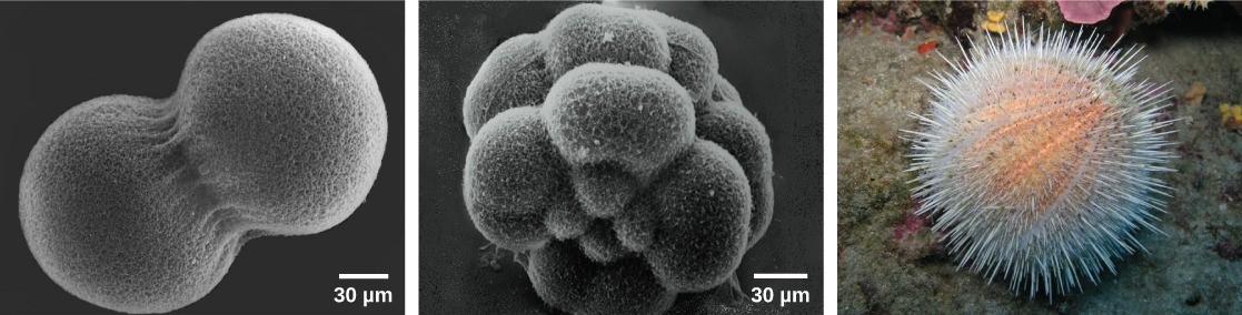
One of the criteria for defining life is growth and development. Multi-cellular organisms grow by multiplying their cells. A human, like every sexually reproducing organism, begins life as a fertilized egg called a zygote (Fig 2.1). In our species, billions of cell divisions subsequently must occur in a controlled manner to produce a complex, multi-cellular human comprising trillions of cells. Thus, the original single-celled zygote is the ancestor of all cells in the body. Once a human is fully grown, cell reproduction is still necessary to repair and regenerate tissues, such as after a scrape or cut or just as part of daily maintenance and repair of tissues. Cell division is a carefully regulated process, and the occasional failure of this regulation can have life-threatening consequences. Single-celled organisms may also use cell division as their method of reproduction.
The Cell Cycle
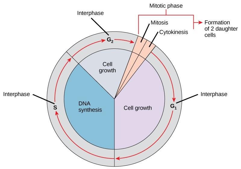
The cell cycle is an ordered series of events involving cell growth and cell division that produces two new daughter cells. Cells on the path to cell division proceed through a series of precisely timed and carefully regulated stages of growth, DNA replication, and nuclear and cytoplasmic division that ultimately produces two identical (clone) cells. The cell cycle has two major phases: the interphase and the mitotic phase (Figure 2.2). During interphase, the cell grows and DNA is replicated. During the mitotic phase, the replicated DNA and cytoplasmic contents are separated, and the cell cytoplasm is typically partitioned by a third process of the cell cycle called cytokinesis. Cytokinesis is defined as a separate process because some cells undergo interphase and mitosis without cytokinesis, resulting in cells with multiple nuclei (multinucleate cells).
Interphase
During interphase, the cell undergoes normal growth processes while also preparing for cell division. For a cell to move from the interphase into the mitotic phase, many internal and external conditions must be met. The three stages of interphase are called G1, S, and G2. The details of these phases vary a bit between prokaryotes and eukaryotes so we will focus on the details of the cell cycle in eukaryotic organisms for now.
G1 Phase (First Gap)
The first interphase stage is called the G1 phase (first gap) because, from a microscopic point of view, little change is visible. However, during the G1 stage, the cell is quite active at the biochemical level. The cell is accumulating the building blocks of DNA and the associated proteins as well as accumulating sufficient energy reserves to complete the task of replicating all of its DNA.
S Phase (Synthesis of DNA)
Throughout the interphase, nuclear DNA remains in a semi-condensed chromatin configuration. In the S phase, DNA replication can proceed through the mechanisms that result in the formation of identical pairs of DNA molecules—sister chromatids—that are firmly attached to each other at the centromere .
G2 Phase (Second Gap)
In the G2 phase, the cell replenishes its energy stores and synthesizes proteins necessary for chromosome manipulation and movement that will take place in mitosis. Some cell organelles are duplicated and the cytoskeleton is dismantled to provide resources for the mitotic phase. There may be additional cell growth during G2. The final preparations for the mitotic phase must be completed before the cell is able to enter the first stage of mitosis.
The Mitotic Phase
The mitotic phase is a multistep process during which the duplicated chromosomes are aligned, separated, and move into two new, identical daughter cells. The first portion of the mitotic phase includes mitosis, which we will learn about in much more detail in the next chapter. The second portion of the mitotic phase (a process separate from and following mitosis) is called cytokinesis—the physical separation of the cytoplasmic components into the two daughter cells.
Mitosis
Mitosis is the process of nuclear division. The identical copies of chromosomes separate into two separate nuclei within a single cell. We will learn much more about this process in the coming chapters and class periods.
Cytokinesis
Cytokinesis, or “cell motion,” is the second main stage of the mitotic phase, during which cell division is completed via the physical separation of the cytoplasmic components into two daughter cells. Cytokinesis can be viewed as a separate phase that may or may not occur following mitosis. While most cells will undergo cytokinesis immediately after mitosis to create two identical daughter cells, other cells undergo mitosis without cytokinesis resulting in cells with multiple nuclei (Figure 2.3). Although the stages of mitosis are similar for most eukaryotes, the process of cytokinesis is quite different for eukaryotes with cell walls, such as plant cells.
Reading Check #1
Which phase of the cell cycle involves DNA replication?
A. G1 phase
B. S phase
C. G2 phase
D. Mitotic phase
G0 Phase (Quiescent phase)
Not all cells adhere to the classic cell-cycle pattern in which a newly formed daughter cell immediately enters the preparatory phases of interphase, closely followed by the mitotic phase and cytokinesis. Cells in the G0 phase are not actively preparing to divide. The cell is in a quiescent (inactive) stage that occurs when cells exit the cell cycle. Some cells enter G0 temporarily due to environmental conditions such as the availability of nutrients or stimulation by growth factors. The cell will remain in this phase until conditions improve or until an external signal triggers the onset of G1. Other cells rarely or never divide, such as mature cardiac muscle and nerve cells, which remain in G0 permanently.
Research Connection: Dr. Mary Dasso
Dr. Mary Dasso is an American biochemist whose research focuses on mechanisms of chromosome segregation. Dr. Dasso’s research team discovered that Ran GTPase, a “protein that helps move other proteins between the nucleus and the rest of the cell”, is critical in spindle assembly and chromosome segregation (NIH, 2019; NIH 2022). Her team also discovered that Ran GTPase performs its functions independently of nuclear transport; this finding directly challenged the status quo, and many people advised her to stop working on the project! Her team’s research changed the scientific community’s understanding of how a chromosome communicates with the rest of the cell (NIH, 2019). Learn more about Dr. Dasso’s past and ongoing research by visiting the Mary Dasso Lab website.
Reading Check #2
During which phase of the cell cycle does cytokinesis occur?
A. G1 phase
B. S phase
C. G2 phase
D. Mitotic phase
Regulation of the Cell Cycle
The length of the cell cycle is highly variable, even within the cells of a single organism. In humans, the frequency of cell turnover ranges from a few hours in early embryonic development to an average of two to five days for epithelial cells and to an entire human lifetime spent in G0 by specialized cells, such as cortical neurons or cardiac muscle cells.
There is also variation in the time that a cell spends in each phase of the cell cycle. When rapidly dividing mammalian cells are grown in a culture (outside the body under optimal growing conditions), the cell cycle length is about 24 hours. In rapidly dividing human cells with a 24-hour cell cycle, the G1 phase lasts approximately 9 hours, the S phase lasts 10 hours, the G2 phase lasts about 4.5 hours, and the M phase lasts approximately 0.5 hours. By comparison, in fertilized eggs (and early embryos) of fruit flies, the cell cycle is completed in about 8 minutes. This is because the nucleus of the fertilized egg divides many times by mitosis but does not go through cytokinesis until a multinucleate “zygote” has been produced, with many nuclei located along the periphery of the cell membrane, thereby shortening the time of the cell division cycle. The timing and proceeding of events in the cell cycle are controlled by mechanisms that are both internal and external to the cell.
Regulation of the Cell Cycle by External Events
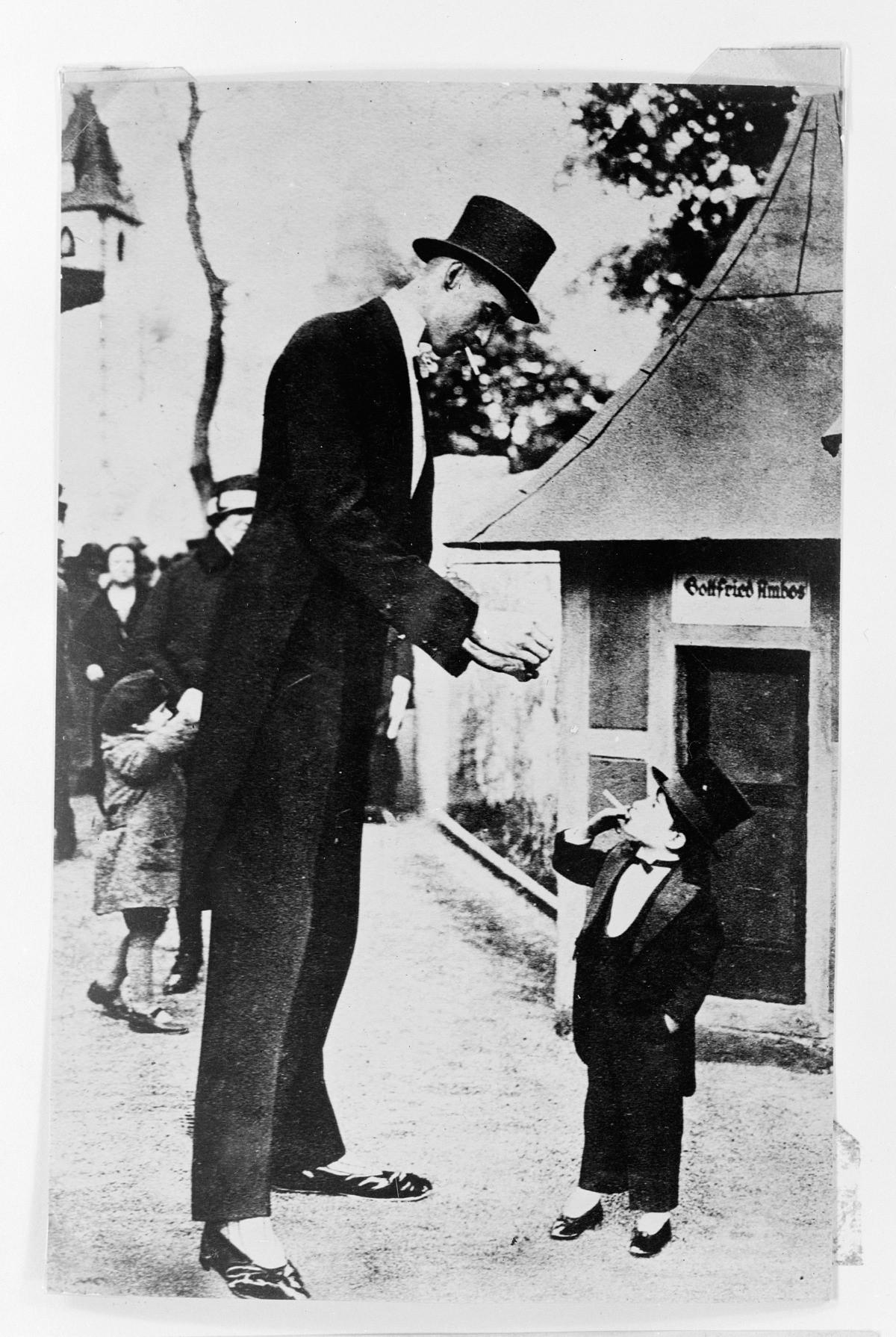
Both the initiation and inhibition of cell division are triggered by events external to the cell when it is about to begin the replication process. An event may be as simple as the death of nearby cells or as sweeping as the release of growth-promoting hormones, such as Human Growth Hormone (HGH). A lack of HGH can inhibit cell division, resulting in dwarfism, whereas too much HGH can result in gigantism (Fig 2.4). Crowding of cells can also inhibit cell division. In contrast, a factor that can initiate cell division is the size of the cell: As a cell grows, it becomes physiologically inefficient due to its decreasing surface-to-volume ratio. The solution to this problem is to divide. Whatever the source of the message, the cell receives the signal, and a series of events within the cell allows it to proceed into interphase. Moving forward from this initiation point, every parameter required during each cell cycle phase must be met, or the cycle cannot progress.
Internal Regulation of the Cell Cycle
It is essential that the daughter cells produced from mitosis are exact duplicates of the parent cell. Mistakes in the duplication or distribution of the chromosomes leads to mutations that may be passed forward to every new cell produced from an abnormal cell. To prevent a compromised cell from continuing to divide, internal control mechanisms operate at three main cell-cycle checkpoints. A checkpoint is one of several points in the eukaryotic cell cycle at which the progression of a cell to the next stage in the cycle can be halted until conditions are favorable. These checkpoints occur near the end of G1, at the G2/M transition, and during metaphase within mitosis (the M checkpoint) (Figure 2.5).
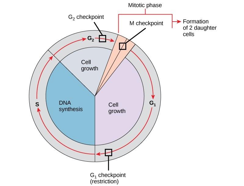
G1 Checkpoint
The G1 checkpoint determines whether conditions are favorable for cell division to proceed. The G1 checkpoint is the point at which the cell irreversibly commits to the cell division process. External influences, such as growth factors, play a large role in carrying the cell past the G1 checkpoint. At the G1 checkpoint, the cell checks to ensure that there are adequate reserves of energy and resources, the cell size is appropriate, and there is no DNA damage. A cell that does not meet all the requirements will not be allowed to progress into the S phase. The cell can halt the cycle and attempt to remedy the problematic condition, or the cell can move into G0 (Quiescent phase) and await further signals when conditions improve.
G2 Checkpoint
The G2 checkpoint bars entry into the mitotic phase if certain conditions are not met. Similar to the G1 checkpoint, cell size and protein reserves are assessed. However, the most important role of the G2 checkpoint is to ensure that all of the chromosomes have been replicated and that the replicated DNA is not damaged. If the checkpoint mechanisms detect problems with the DNA, the cell cycle is halted, and the cell attempts to either complete DNA replication or repair the damaged DNA.
M Checkpoint
The M checkpoint occurs near the end of the third stage (metaphase) of mitosis. Recall that in M phase, identical copies of chromosomes separate into two separate nuclei. The equal distribution of chromosomes is vital to ensure each daughter cell has the correct number of chromosomes. The acquisition or loss of chromosomes results in the formation of daughter cells that are incorrectly coded. The M checkpoint determines whether all the sister (duplicated) chromatids are prepared to migrate appropriately.
Reading Check #3
What is the role of the G2 checkpoint in the cell cycle?
A. Assess cell size and protein reserves.
B. Ensure adequate energy and resource reserves.
C. Verify that all chromosomes have been replicated and are undamaged.
D. Determine whether all sister chromatids are prepared to migrate.
Regulator Molecules of the Cell Cycle
In addition to the internally controlled checkpoints, there are two groups of intracellular molecules that regulate the cell cycle. These regulatory molecules either promote the progress of the cell to the next phase (positive regulation) or halt the cycle (negative regulation). Regulator molecules may act individually or influence the activity or production of other regulatory proteins. Therefore, the failure of a single regulator may have almost no effect on the cell cycle, especially if more than one mechanism controls the same event. However, the effect of a deficient or non-functioning regulator can be wide-ranging and possibly fatal to the cell if multiple processes are affected.
Positive Regulation of the Cell Cycle
Two protein groups, cyclins and cyclin-dependent kinases (Cdks), are positive regulators. They are responsible for the progress of the cell through the various checkpoints. The levels of the four cyclin proteins fluctuate throughout the cell cycle in a predictable pattern (Fig 2.6). Both external and internal signals trigger increases in the concentration of cyclin proteins. After the cell moves to the next stage of the cell cycle, the active cyclins in the previous stage are degraded by cytoplasmic enzymes, producing the cycles as shown in Figure 2.6.
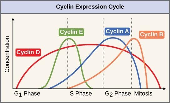
Cyclins regulate the cell cycle only when tightly bound to a group of enzymes called Cdks (which stands for cyclin-dependent kinases). To be fully active, the Cdk/cyclin complex must also be phosphorylated (meaning a phosphate group is attached) in specific locations to activate the complex. Cdks belong to a class of enzymes called kinases. Kinases is the name given to enzymes that phosphorylate (i.e., add a phosphate group to) other proteins. Phosphorylation activates the target protein by changing its shape. The proteins phosphorylated by Cdks are involved in advancing the cell to the next phase. The levels of Cdks are relatively stable throughout the cell cycle; however, the concentrations of cyclin fluctuate and determine when Cdk/cyclin complexes form. The different cyclins and Cdks bind at specific points in the cell cycle and thus regulate different checkpoints.
Because the cyclic fluctuations of cyclin levels are based mainly on the timing of the cell cycle and not on specific events, regulation of the cell cycle usually occurs by either the Cdk molecules alone or the Cdk/cyclin complexes. Without a specific concentration of fully activated cyclin/Cdk complexes, the cell cycle cannot proceed through the checkpoints.
Although the cyclins are the main regulatory molecules that determine the forward momentum of the cell cycle, there are several other mechanisms that fine-tune the progress of the cycle with negative rather than positive effects. These mechanisms essentially block the progression of the cell cycle until problematic conditions are resolved. Molecules that prevent the full activation of Cdks are called Cdk inhibitors. Many of these inhibitor molecules directly or indirectly monitor a particular cell-cycle event. The block placed on Cdks by inhibitor molecules will not be removed until the specific event that the inhibitor monitors is completed.
Negative Regulation of the Cell Cycle
The second group of cell-cycle regulatory molecules is negative regulators, which stop the cell cycle. Remember that in positive regulation, active molecules cause the cycle to progress.
The best-understood negative regulatory molecules are retinoblastoma protein (Rb), p53, and p21. Much of what is known about cell-cycle regulation comes from research conducted with cells that have lost regulatory control. All three of these regulatory proteins were discovered to be damaged or non-functional in cells that had begun to replicate uncontrollably (i.e., become cancerous). In each case, the main cause of the unchecked progress through the cell cycle was a faulty copy of the regulatory protein.
Rb, p53, and p21 act primarily at the G1 checkpoint. p53 is a multi-functional protein that has a major impact on the commitment of a cell to division because it acts when there is damaged DNA in cells that are undergoing the preparatory processes during G1. If damaged DNA is detected, p53 halts the cell cycle and then recruits specific enzymes to repair the DNA. If the DNA cannot be repaired, p53 can trigger apoptosis, or cell suicide, to prevent the duplication of damaged chromosomes. As p53 levels rise, the production of p21 is triggered. p21 enforces the halt in the cycle dictated by p53 by binding to and inhibiting the activity of the Cdk/cyclin complexes. As a cell is exposed to more stress, higher levels of p53 and p21 accumulate, making it less likely that the cell will move into the S phase.
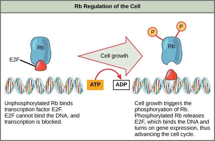
Rb, which largely monitors cell size, exerts its regulatory influence on other positive regulator proteins. In the active, dephosphorylated state, Rb binds to proteins called transcription factors, most commonly E2F (Fig 2.7). Transcription factors “turn on” specific genes, allowing the production of proteins encoded by that gene.
When Rb is bound to E2F, the production of proteins necessary for the G1/S transition is blocked. As the cell increases in size, Rb is slowly phosphorylated until it becomes inactivated. Rb releases E2F, which can now turn on the gene that produces the transition protein, and this particular block is removed. For the cell to move past each of the checkpoints, all positive regulators must be “turned on,” and all negative regulators must be “turned off.”
Rb and other proteins that negatively regulate the cell cycle are called tumor suppressors. Why do you think the name tumor suppressor might be appropriate for these proteins?
Reading Check #4
What would be a potential consequence of a mutation at the p53 gene?
A. Mutant p53 proteins would not function as tumor suppressors.
B. Mutant p53 proteins would no longer function as an oncogene.
C. Mutant p53 proteins will prevent the cell from entering S phase.
D. All of the above would be potential consequences.
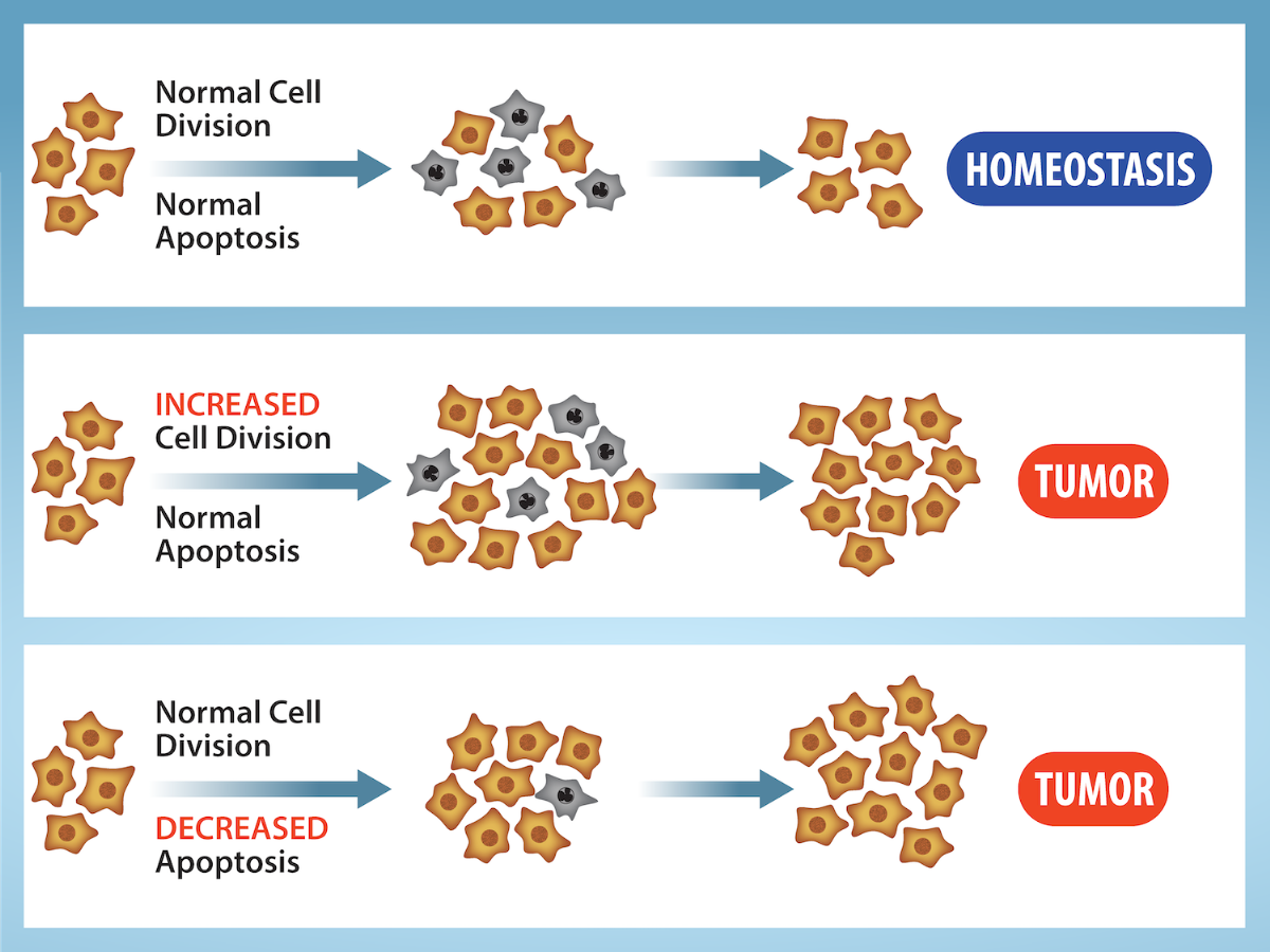
Cancer and the Cell Cycle
Cancer comprises many different diseases caused by a common mechanism: uncontrolled cell growth. Despite the redundancy and overlapping levels of cell-cycle control, errors do occur. One of the critical processes monitored by the cell-cycle checkpoint surveillance mechanism is the proper replication of DNA during the S phase. Even when all of the cell-cycle controls are fully functional, a small percentage of replication errors (mutations) will be passed on to the daughter cells. If changes to the DNA sequence occur within a coding portion of a gene and are not corrected, a gene mutation results. Cancers start when a gene mutation gives rise to a faulty protein that plays a key role in cell reproduction.
The change in the cell that results from the malformed protein may be minor: perhaps a slight delay in the binding of Cdk to cyclin or an Rb protein that detaches from its target DNA while still phosphorylated. Even minor mistakes, however, may allow subsequent mistakes to occur more readily. Over and over, small uncorrected errors are passed from the parent cell to the daughter cells and amplified as each generation produces more non-functional proteins from uncorrected DNA damage. Eventually, the pace of the cell cycle speeds up as the effectiveness of the control and repair mechanisms decreases. Uncontrolled growth of the mutated cells outpaces the growth of normal cells in the area, and a tumor can result (Fig 2.8).
Proto-oncogenes
The genes that code for the positive cell-cycle regulators are called proto-oncogenes. Proto-oncogenes are normal genes that, when mutated in certain ways, become oncogenes—genes that cause a cell to become cancerous. Consider what might happen to the cell cycle in a cell with a recent mutation in a proto-oncogene. In most instances, the alteration of the DNA sequence will result in a less functional (or non-functional) protein. The result is detrimental to the cell and will likely prevent the cell from completing the cell cycle; however, the organism is not harmed because the mutation will not be carried forward since the cell is not reproducing itself. If a cell cannot reproduce, the mutation is not propagated, and the damage is minimal. Occasionally, however, a mutation in a proto-oncogene causes a change that increases the activity of a positive regulator. For example, a mutation that allows Cdk to be activated without being partnered with cyclin could push the cell cycle past a checkpoint before all of the required conditions are met. If the atypical daughter cells are able to undergo further cell divisions, subsequent generations of cells may accumulate even more mutations, some possibly in additional genes that regulate the cell cycle.
Tumor Suppressor Genes
Like proto-oncogenes, many of the negative cell-cycle regulatory proteins were discovered in cells that had become cancerous. Tumor suppressor genes are genes that code for negative regulator proteins, the type of regulators that, when activated, can prevent the cell from undergoing uncontrolled division. The collective function of the best-understood tumor suppressor gene proteins, Rb, p53, and p21, is to put up a roadblock to cell-cycle progression until certain events are completed. A cell that carries a mutated form of a negative regulator might not be able to halt the cell cycle if there is a problem. Tumor suppressors are similar to brakes in a vehicle: Malfunctioning brakes can contribute to a car crash! Mutated p53 genes have been identified in more than 50% of all human tumor cells. This discovery is not surprising in light of the multiple roles that the p53 protein plays at the G1 checkpoint. A cell with a faulty p53 may fail to detect errors present in the genomic DNA (Fig 2.9). Even if a partially functional p53 does identify the mutations, it may no longer be able to signal the necessary DNA repair enzymes. Either way, damaged DNA will remain uncorrected. At this point, a functional p53 will deem the cell unsalvageable and trigger programmed cell death (apoptosis). The damaged version of p53 found in cancer cells, however, cannot trigger apoptosis.
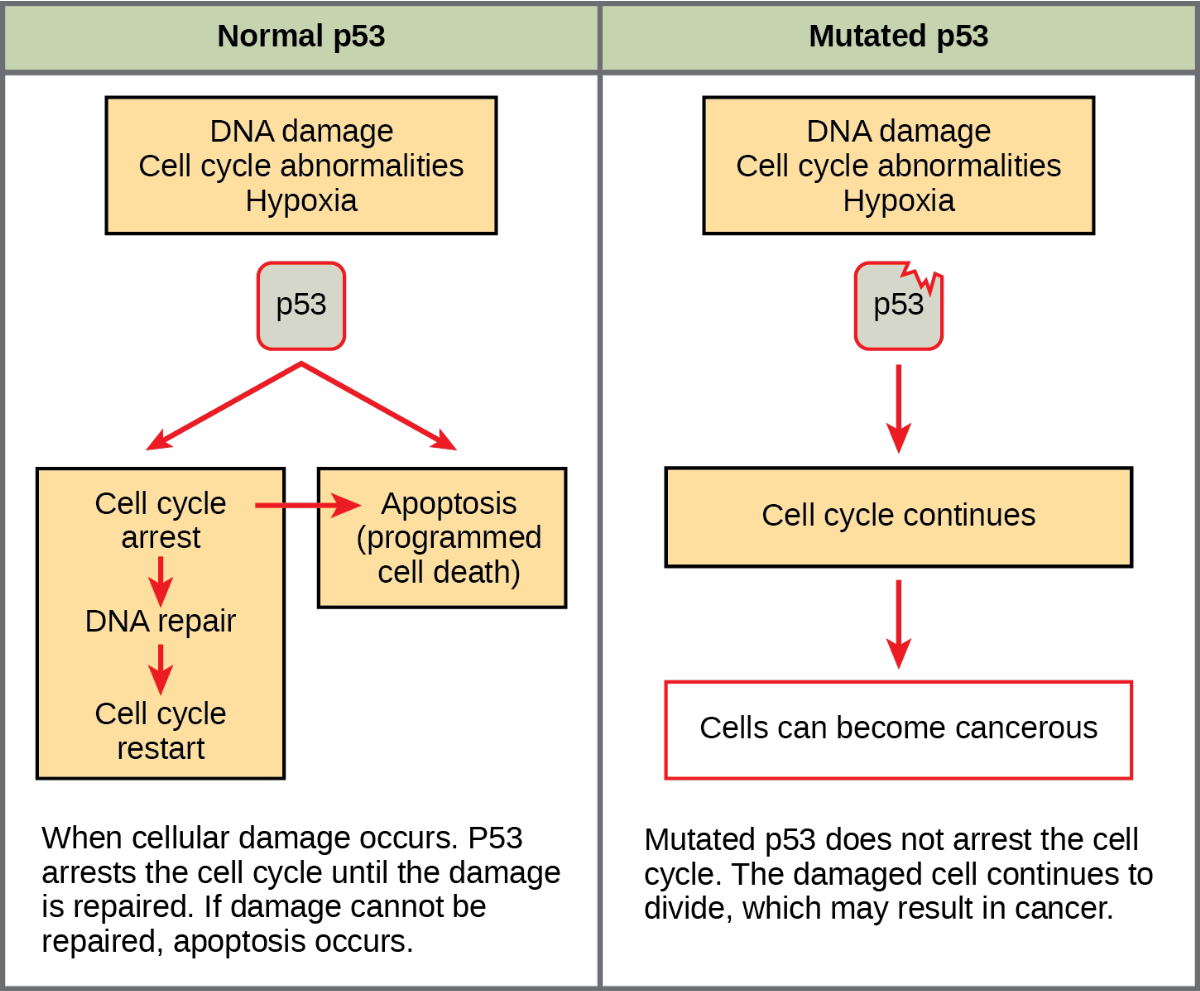
The loss of p53 function has other repercussions for the cell cycle. Mutated p53 might lose its ability to trigger p21 production. Without adequate levels of p21, there is no effective block on Cdk activation. Essentially, without a fully functional p53, the G1 checkpoint is severely compromised and the cell proceeds directly from G1 to S regardless of internal and external conditions. At the completion of this shortened cell cycle, two daughter cells are produced that have inherited the mutated p53 gene. Given the non-optimal conditions under which the parent cell reproduced, it is likely that the daughter cells will have acquired other mutations in addition to the faulty tumor-suppressor gene. Cells such as these daughter cells quickly accumulate both oncogenes and non-functional tumor-suppressor genes. Again, the result is tumor growth.
Reading Check #5
Which types of genes, when mutated, become oncogenes and cause a cell to become cancerous?
A. Tumor suppressor genes
B. Proto-oncogenes
C. Negative regulator genes
D. p53 and p21
References and Acknowledgements
Adapted from Boundless. (2023). General Biology. LibreTexts. Retrieved from https://bio.libretexts.org/Bookshelves/Introductory_and_General_Biology/Book%3A_General_Biology_(Boundless).
