
18 Chapter 18. Form Follows Function
Anastasia Chouvalova and Joshua Reid
Learning Objectives:
By the end of this section, students will be able to:
18.1 Explain the biological idea of “form follows function” and support your explanation with specific examples.
18.2 Use the concept of “form follows function” to explain why different cell/tissue types look different and give examples.
Form follows Function
One of the overarching themes of biology is that form follows function; how something is arranged allows it to perform a specific job. We see this at all levels in the hierarchy of biological organization from atoms up to the biosphere.
Molecular level – proteins
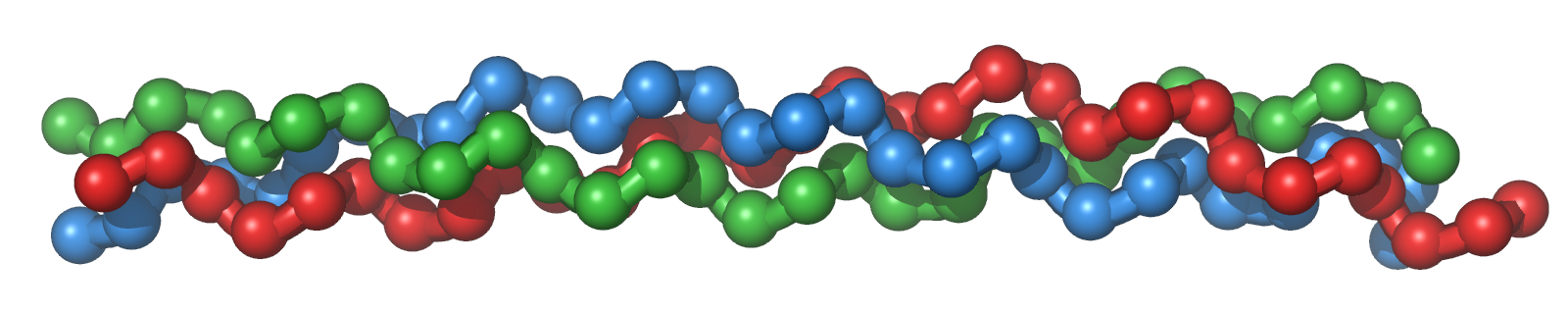
The shape (structure) of a protein determines its function and the function of a protein often directs how it is shaped three-dimensionally (i.e., tertiary structure). For example, there are two basic shapes for proteins: fibrous and globular (round). Collagen is an example of a fibrous protein (Figure 18.1) that gives strength to our skin to prevent it from tearing and for this purpose, it is shaped like a rope. Fibrous proteins are structural proteins because they help give shape to and support the skin. This is an example of form follows function at the molecular level of proteins.
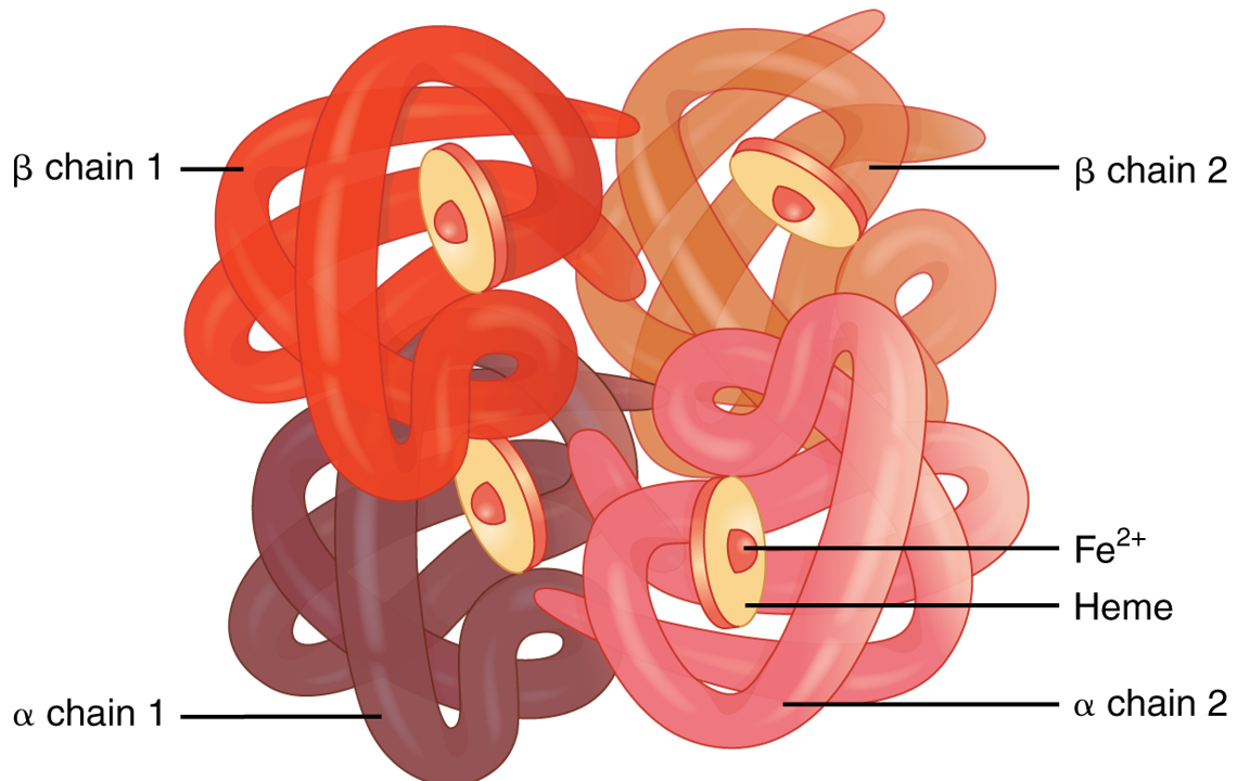
The function of globular proteins, such as hemoglobin (Figure 18.2), is to transport oxygen in the blood so that all cells of the body can receive oxygen to perform metabolic processes. Other examples of globular proteins that have different functions are enzymes (catalyze or speed up chemical reactions in the body) and plasma membrane proteins (can transport substances across the cell membrane, play a role in cell communication, act as enzymes, or help identify the cell to the rest of the body).
Cellular level – skeletal muscle cells
The structure of skeletal muscle cells allows them to have the function of contraction, which allows us to move. But how a muscle is used also affects its structure and function – an overused muscle will often hypertrophy or in other words, have a larger cross-sectional area. A cell can change its shape and structure, and even its volume, if demands drastically change (e.g., more force production is needed).
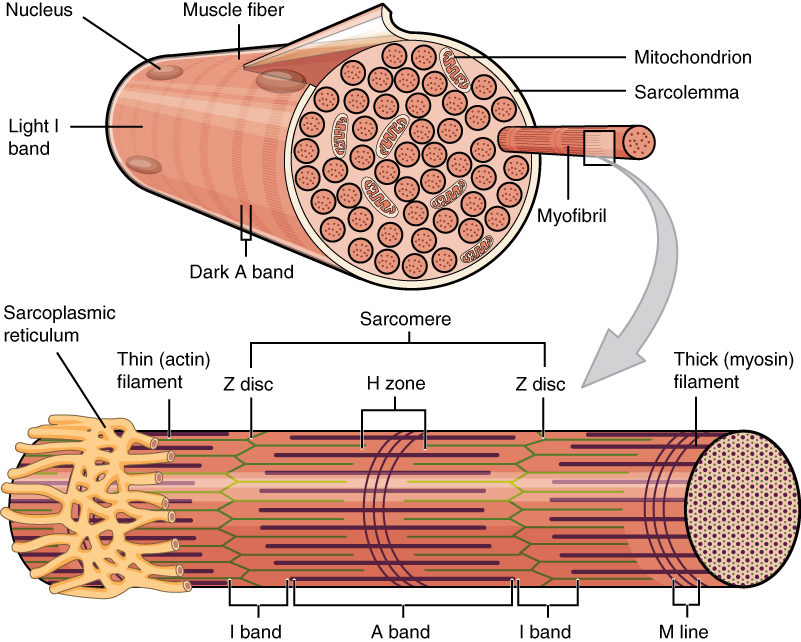
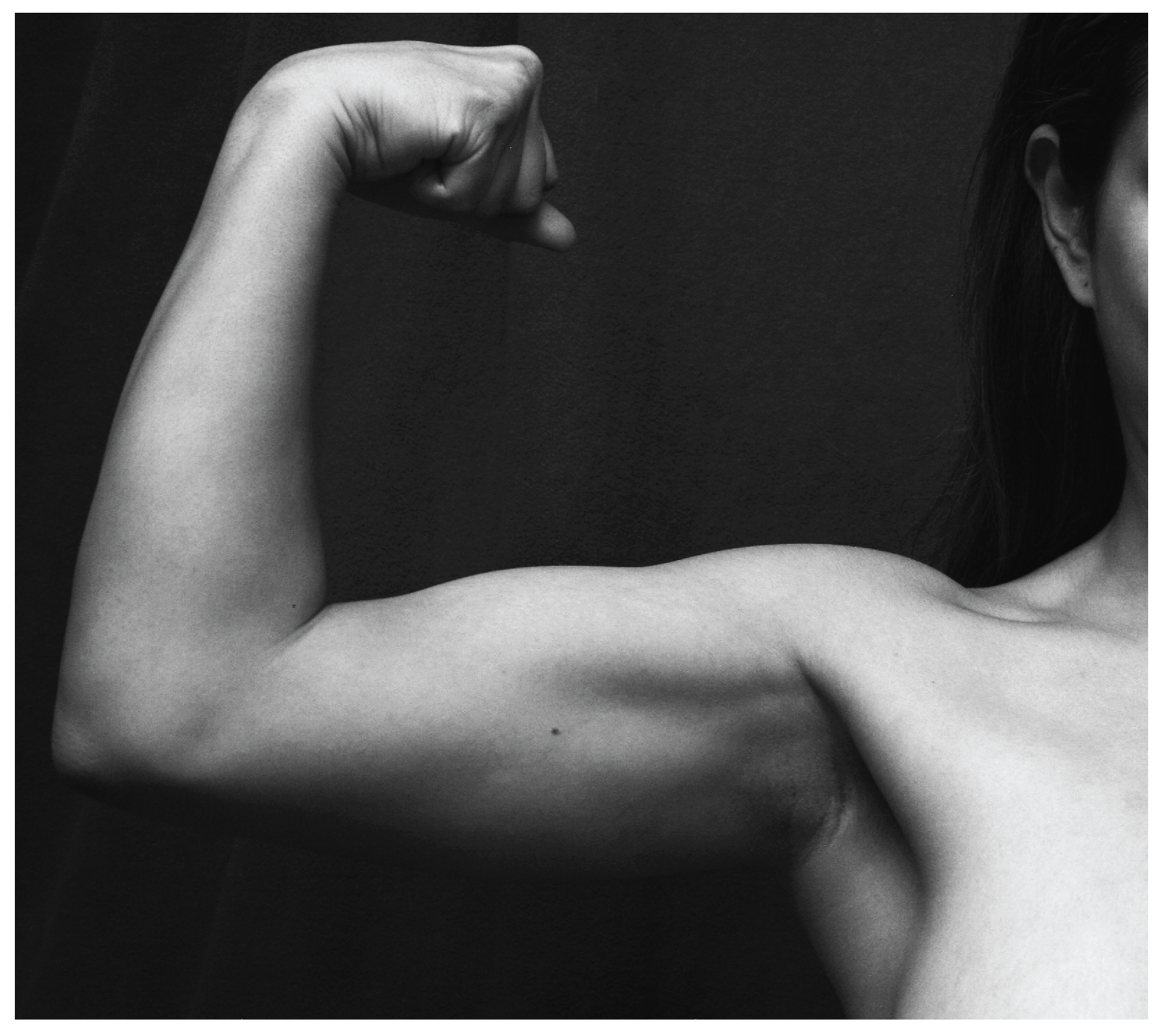
Skeletal muscle cells that make up your biceps brachii muscle are attached to both ends of the humerus bone by tendons and are packed full of contractile proteins (actin and myosin) (Figure 18.3). When the contractile proteins contract, they shorten the muscle cell, which then pulls on the ends of the humerus and allows you to flex your forearm (Figure 18.4).
Individual Level
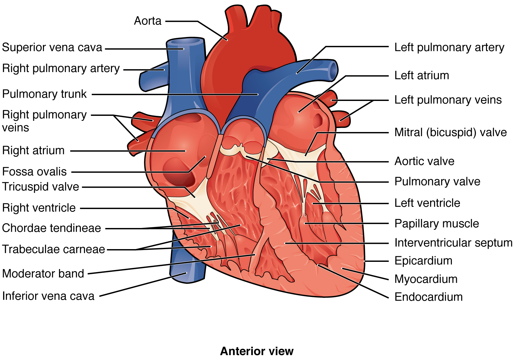
In studying humans, anatomy is the study of the structure of the body (ex- where the quadriceps muscle is located) and physiology is the study of how the body functions (ex- how the quadriceps muscle contracts). Let’s take a look at the function and anatomy of the heart, which pumps blood to the entire body. The heart consists of four hollow chambers (atria and ventricles) and is made of cardiac muscle cells (Figure 18.5). This structure allows the heart to have the function of pumping blood around the body. However, if the structure of the heart changes (ex- some of the heart chambers become stretched out or dilated), then the heart’s function decreases as the heart can no longer pump as much blood, which will eventually cause congestive heart failure.
On the other hand, if one of the heart chambers loses functionality, it will affect that chamber’s shape. For example, if the left ventricle stops pumping blood to the left atrium, its walls will thicken, leading to left ventricular hypertrophy. This is an example of form follows function, or more specifically, impaired form follows impaired function, in cardiac physiology.
Ecosystem level
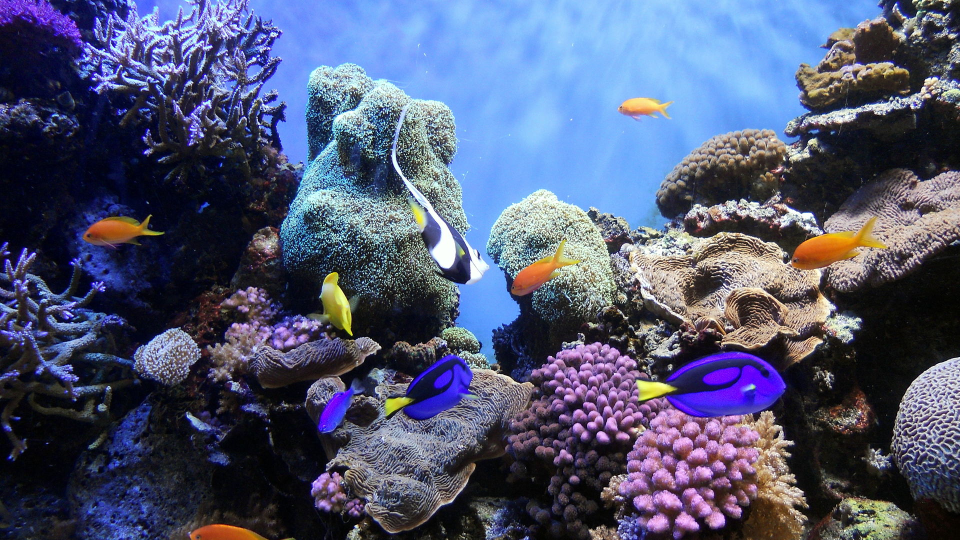
An ecosystem consists of a community of all the different species living in a particular geographic area as well as all of the nonliving components (ex- water, sand, light, oxygen). If we look at the structure of a coral reef ecosystem, we see that the corals, which are the foundation species, provide protection and habitat for other species (Figure 18.6). The coral reef protects other species, such as fish, from ocean waves and currents and gives them a place to hide from predators. If the coral reef’s protective function is disrupted, the form of the overall ecosystem will change – the food web and food chain inherent to that ecosystem will be thrown off balance. For example, if the coral reef’s structural integrity is damaged, fish at lower trophic levels will die since they are more vulnerable to the ocean waves, currents, and predators. This will deprive fish at higher trophic levels of prey. This is an example of form follows function at the broader ecosystem level. In this case, impaired function at lower trophic levels impacts the integrity of higher trophic levels.
Reading Question #1
What is meant by the phrase “form follows function”? Select the best answer.
A. How an organism or part of an organism is shaped is related to its purpose and use.
B. The shape and structure of an organism assists in its fitness and performance.
C. How an organism or part of an organism is structured produces its purpose and use.
D. The functionality of an organism assists in its fitness and performance.
Reading Question #2
Of the following, which is the best example of the phrase “form follows function” in the physiological context? (Hint: What happens if you lift greater weights?)
A. A weightlifter notices greater muscle mass in his arm muscles, which allows him to increase his load and do more repetitions of the increased load.
B. A weightlifter notices greater cross-sectional area of his arm muscles, which allows him to increase his load and do more repetitions of the increased load.
C. A weightlifter is increasing his load and doing more repetitions of the increased load, resulting in muscle hypertrophy and greater muscle mass.
D. A weightlifter is decreasing his load and doing less repetitions of the decreased load, resulting in muscle hypertrophy and greater muscle mass.
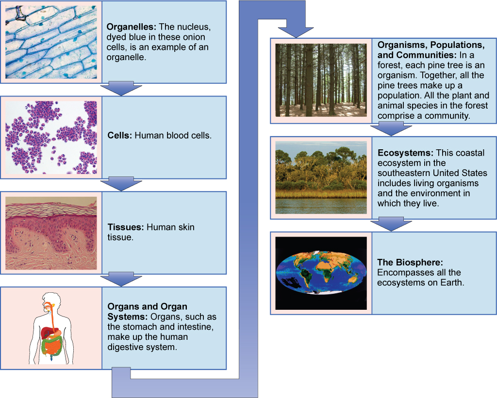
Biological Levels of Organization
Reading Question #3
Of the following, identify the correct statement(s). Select all that apply.
A. Biospheres exist within organs which in turn, exist within organ systems.
B. Ecosystems are compromised of communities which in turn, are compromised of different populations.
C. Various cells exist within tissues which in turn, comprise organs.
D. Organs comprise cells which in turn, comprise tissues.
E. A community is made up of only animal species and excludes plant species.
F. Organs are comprised of various tissues which in turn, are made up of different cells.
G. The biosphere contains ecosystems which in turn, are made up of communities of populations.
Human Tissue Types
The term tissue is used to describe a group of similar cells found together in the body that act together to perform specific functions. From the evolutionary perspective, tissues appear in more complex organisms.
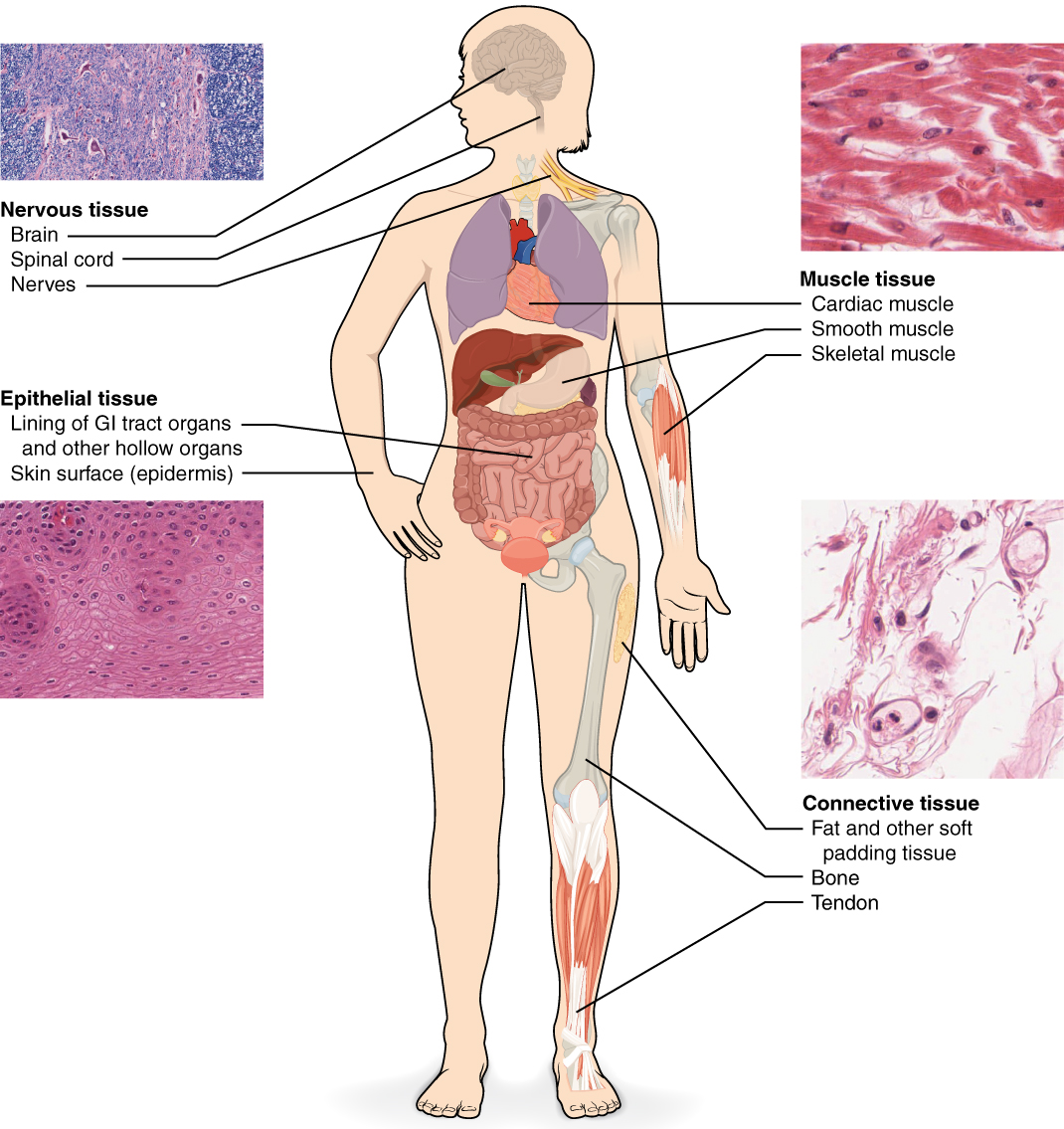
Although there are many types of cells in the human body, they are organized into four categories of tissues: epithelial, connective, muscle, and nervous. Each of these categories is characterized by specific functions that contribute to the overall health and maintenance of the body. A disruption of the structure of a tissue is a sign of injury or disease. Such changes can be detected through histology, the microscopic study of tissue appearance, organization, and function.
The Four Types of Tissues
Epithelial tissue, also referred to as epithelium, refers to the sheets of cells that cover exterior surfaces of the body, line internal cavities and passageways, and form certain glands. Examples of epithelial tissue include skin, mucous membranes, endocrine glands, and sweat glands. Connective tissue, as its name implies, binds the cells and organs of the body together and functions in the protection, support, and integration of all parts of the body. Connective tissue is diverse and includes bone, tendons, ligaments, cartilage, fat, and blood. Muscle tissue is excitable, responding to stimulation and contracting to provide movement, and occurs as three major types: skeletal (voluntary) muscle, smooth muscle, and cardiac muscle in the heart. Nervous tissue is also excitable, allowing the propagation of electrochemical signals in the form of nerve impulses that communicate between different regions of the body (Figure 18.8).
The next level of organization is the organ, where two or more types of tissues come together to perform specific functions. Just as knowing the structure and function of cells helps you in your study of tissues, knowledge of tissues will help you understand how organs function.
Reading Question #4
Of the four tissue types, which are excitable? Select all that apply.
A. Muscle
B. Epithelial
C. Nervous
D. Connective
Reading Question #5
Of the four tissue types, which one is used to form a protective barrier?
A. Nervous
B. Connective
C. Muscle
D. Epithelial
References
Adapted from Clark, M.A., Douglas, M., and Choi, J. (2018). Biology 2e. OpenStax. Retrieved from https://openstax.org/books/biology-2e/pages/1-2-themes-and-concepts-of-biology?query=%22organ%20system%22&target=%7B%22type%22%3A%22search%22%2C%22index%22%3A0%7D#fs-id2155753
Barrickman, N., Bell, K., and Cowan, C. (n.d.) Human Biology. Pressbooks. Retrieved from https://slcc.pressbooks.pub/humanbiology/chapter/chapter-12-organ-systems-of-the-human-body/
